Our Formulas - The Clear Ocluvet Difference
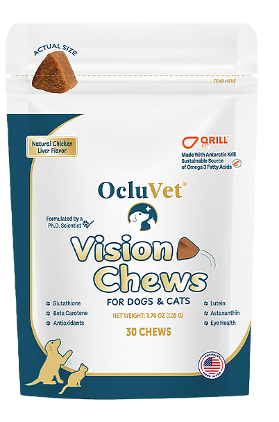
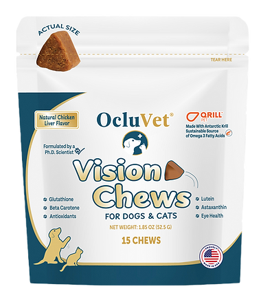
Introducing OcluVet® Vision Chews
Made With QRILL™ PET
Antarctic Krill Which Benefits:
Skin and Coat -Fat is very important for healthy skin and coat in pets. A regular diet based on essential fatty acids like those found in krill is essential to keep the skin barrier fit and the coat shiny.
Liver - Omega-3s and choline improve and maintain a healthy liver function, and can aid in the proper metabolism of fat.
Joints - Krill omega-3s have anti-inflammatory properties and can contribute to reducing inflammation and thereby joint pain caused by wear and tear due to aging.
Brain - Omega-3s and choline from krill are essential in supporting brain development, the learning process, the nerve transmitters and affect the overall mental well-being of pets.
Heart - Omega-3s are important for a healthy heart and can help lower blood pressure and prevent dangerous blood clots that could be damaging to the heart.
Immune system - A healthy, balanced diet that includes omega-3s and astaxanthin can support and enhance the immune system of cats and dogs of all ages, making immune cells more flexible and resistant.
OcluVet® Patented Eye Drops
For Pets Contain:
N-Acetyl-L-Carnosine (NAC 2%) - a naturally occurring molecule clinically proven to help slow, reduce and even reverse lens opacity of immature cataracts, with an 80+% success rate when treated early on.
L-Carnosine - Neutralizes free radicals.
L-Gluatathione - A powerful antioxidant that works in synergy with NAC. This is important for protecting protein, DNA repair & only in our formula.
Cysteine and Riboflavin - Recycles the eye's endogenous glutathione to its active form
Ascorbic acid - Vitamin C which also helps with the metabolism of glutathione
Taurine - An antioxidant with detoxifying activity that helps stabilize cell membranes


These photographs are of a U.S. study participant. The photo on the left is pretreatment and the photo on the right is at 8 weeks post treatment. The supervising veterinarian noted that at the 6 and 8 week exams the retinal vessels were visible where previously they could not be seen. This is clearly demonstrated in the photos.


